The OSU Biomedical Imaging Center provides superior screening and diagnostic imagery with Prisma and Vida 3T MRIs. Our world-class technology offers shorter scan times which result in improved diagnosis and care plans for physicians and their patients. With Prisma and Vida scanners, we support virtually all neurological and musculoskeletal imaging, both with and without gadolinium contrast. Our outpatient clinic is staffed with highly-trained experts and offers flexible scheduling and convenient parking.

OSU Biomedical Imaging Center offers multiple MRI modalities:

  • Breast
  • Body
  • MR Angiography (MRA)
  • Musculoskeletal/Orthopedic
  • Neurological (Brain and Spine)

Additional Benefits:

  • Competitive self-pay prices for patients who are uninsured or have a high deductible plan.
  • Convenient parking available just feet away from the clinic entrance.
  • The price for non-contrast MRI scans is $425; and $525 for contrast MRI scans. All prices include the radiologist reading-fee.
  • MRI Scans available Monday through Friday from 7:30 a.m. – 11 p.m. to accommodate patients’ busy schedules.
  • Offering “Fed & Wrapped” non-sedated pediatric MRI from birth through adolescence.
  • Offering two 3T MRIs: a Siemens PRISMA and a Siemens Vida Wide-Bore Scanner allowing for rapid scheduling and to accommodate patients who struggle with claustrophobia or who need extra space (weight limit of 550lb).
  • Our clinic will obtain all necessary insurance authorizations.
  • Siemens Deep Resolve Boost technology accelerates image collection, resulting in exceptional images often in half the time of other scanners.

The completed MRI Order form can be faxed to: 539-325-6569. Please call 539-325-6560 with any questions.
About MRI Scans

Because of the strong magnetic field used during the exam, certain conditions may prevent you from having the procedure. When scheduling your appointment, our staff will review any restrictions or conditions that may apply.

What to expect during an MRI scan

  • The duration of the procedure varies but average length is 45 minutes to one hour per area scanned.
  • You must lie still during scan. Depending on the area of the body that is being examined, you may be asked to hold your breath for up to 30 seconds.
  • MRI scans can have intermittent noises that are loud, but you will be provided with earplugs or headphones to minimize the noise during the procedure.
  • The technologist will be able to hear and speak with you throughout the exam. They will also provide you with an alarm button to alert them to any discomfort you may experience at any point during the MRI exam.
  • Some MRI exams require an injection of intravenous MRI contrast. Inform the technologist if you experience any discomfort during the injection.

At the OSU Biomedical Imaging Center, it is our goal to provide both cutting-edge imaging and exceptional patient care. We simplify the ordering and scheduling process for you, your staff and patients. After receiving a physician’s faxed order, our front desk staff will facilitate obtaining any necessary insurance authorizations. We then call to pre-register your patient and schedule their MRI, usually within two business days. Upon arriving at our facility, patients will access convenient parking that is only a few steps from our clinic door.

MRI Features

The OSU Biomedical Imaging Center houses two MRIs: A Siemens Magnetom Prisma 3T MRI, and a Siemens Magnetom Vida 3T large-bore MRI. Both scanners include advanced gradient and radio frequency (RF) transmit subsystems as well as a scalable 64-channel digital MRI receivers, and technology supporting massively parallel fMRI with advanced techniques such as sensitivity encoding (SENSE/iPAT), multi-slice/multiband imaging and CAIPIRINHA (Controlled Aliasing In Parallel Imaging Results IN Higher Acceleration). A wide selection of RF multi-channel, receive-only surface coil arrays for imaging human brains, spinal cords, and peripheral body structures are available. This includes system-standard 20-, 32-, and 64-channel receive-only brain arrays, 32-channel spinal array coils, multiple 18-channel body/peripheral flex coils, 16-channel shoulder coil, 18-channel knee coil, and 16-channel NeoCoil breast coil. Both systems have wireless participant physiological monitoring for cardiac, respiration, and blood oxygenation/pulse measurement. The Vida is also equipped with Siemens Deep Resolve Gain/Sharp/Boost AI reconstruction engines.

The Center is also equipped with the auxiliary research equipment one expects in a state-of-the-art neuroimaging research center. This includes:

  • Real-time participant physiological monitoring for cardiac, respiration, and blood oxygenation/pulse measurement.
  • Cambridge Systems MR-compatible high-resolution LCD screens for viewing visual stimuli inside the magnets.
  • Optoacoustics active noise cancelling audio systems that greatly reduce scanner-related sound, allowing for improved auditory stimulus delivery and voice recording inside the scanner.
  • MR-compatible Current Designs fiberoptic button boxes and scroll wheels for collecting participant responses.
  • Framewise Integrated Real-time MRI Monitoring (FIRMM) – a real-time image processing subsystem allowing for the online monitoring of functional neuroimaging data as they are acquired.
  • The scanners are also equipped with MR-compatible MRC 12M-i video cameras with integrated infrared LED that when mounted on the head coil provide excellent visualization of a participant’s face during scanning, even in the dark.
  • The Center also provides a suite of software tools, including PsychoPy and EPrime for stimulus delivery and OpenNFT for real-time neurofeedback training applications.

Hardesty Center for Clinical Research and Neuroscience,

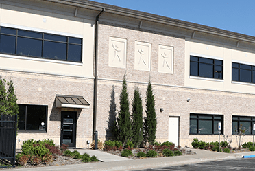
The Hardesty Center for Clinical Research and Neuroscience, which houses the OSU Biomedical Imaging Center, provides state-of-the-art, research infrastructure unmatched in Oklahoma. The 49,000-square-foot research complex houses bioimaging services and a clinical trials unit, along with dedicated faculty and student office space that support biomedical, clinical, and population health research initiatives. The center’s creation is made possible through a gift by the Hardesty Family Foundation and underpins their ongoing commitment to impactful solutions that aid Tulsans for generations.

Early detection just got easier

Definitive answers you need for important breast health decisions are faster and easier with breast MRI services at the OSU Biomedical Imaging Center.

Breast MRI can be a screening procedure for women ages 40–70 with dense breast tissue and who have a low to moderate lifetime breast cancer risk. Research shows that Breast MRI has the highest cancer detection rate of all types of breast imaging across all breast density categories. Breast MRI is most effective when used in conjunction with a mammogram.

Breast MRI can also be a diagnostic tool used for a variety of reasons:

Determining the extent of previously detected breast cancer.
Evaluating abnormalities detected by mammography.
Identifying cancer in women who have implants or scar tissue.
Determining whether lumpectomy or mastectomy would be more effective.
Determining whether breast cancer has spread into the chest wall.
Breast MRIs require an order from your physician and may be covered by your insurance.

Mammography:

Sensitivity is limited by both breast tissue density and complexity.
Uses ionizing radiation and tissue compression to create images of your breast.

Breast MRI:

Sensitivity is not limited by breast tissue density or complexity.
Uses strong magnets to make very detailed images of your breast.